Nobel in Chemistry Awarded for Mapping the Innermost Secrets of Life
Monday, October 13th, 2014October 13, 2014
Two Americans and a German have won the 2014 Nobel Prize in chemistry for their “ground-breaking” efforts in developing ways to peer more closely at the inner workings of living cells using optical (light-bending) microscopes than scientists had thought possible. The winners, all physical chemists, are William E. Moerner of Stanford University in Stanford, California, and Eric Betzig of the Janelia Research Campus at the Howard Hughes Medical Institute in Ashburn, Virginia; and Stefan W. Hell of the Max Planck Institute for Biophysical Chemistry in Göttingen, and the German Cancer Research Center in Heidelberg, both in Germany. The scientists were honored “for the development of super-resolved fluorescence microscopy,” which, the Nobel Committee said, is producing “new knowledge of greatest benefit to mankind … on a daily basis.” They will share the $1.1-million prize equally.
The scientists, working independently, turned the microscope into a nanoscope–a tool for seeing “the pathways of individual molecules inside living cells, according to the Nobel Committee. “They can see how molecules create synapses [bridges] between nerve cells in the brain; they can track proteins involved in Parkinson’s, Alzheimer’s and Huntington’s diseases as they aggregate [clump]; they follow individual proteins in fertilized eggs as these divide into embryos.” Other types of microscopy kill living cells.

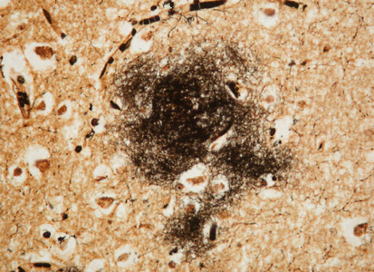
Fluorescence microscopy allows researchers to track the proteins involved in Alzheimer’s disease as they clump. In this photomicrograph, the round, yellow-brown spots, or plaques, are made up of abnormal amyloid protein. (© Jan Naslund, Karolinska Institute, Stockholm, Sweden)
In fluorescence microscopy, cells or parts of cells are stained with green fluorescent protein (GFP), a naturally occurring molecule that glows green when exposed to blue or ultraviolet light. The light is typically captured by a digital camera or another detector connected to the microscope. By using GFP effectively and manipulating the captured images, scientists can identify tiny details within a cell. Since the 1870′s, scientists had believed that optical microscopes would never be able to visualize anything smaller than 0.2 micrometers, a limit based on the wavelength of light. Using fluorescence microscopy, researchers can visualize objects that are at least three times as small as that. Under certain circumstances, the resolution (ability to produce a clear image) may be even greater.
Additional World Book articles:
- Ernst Abbe
- Fluorescence
- Nobel Prizes in chemistry (table)
- Nobel Prizes (2008) (a Back in Time article)


